Partial dentures offer a reliable and effective solution for patients with edentulous areas. When a partial involves treating a patient’s aesthetic zone, communication and alignment among the patient, the clinician, and the lab are crucial for successful treatment. Today, digital dentistry allows us to capture accurate and consistent scans of not only teeth, but also edentulous sites and soft tissue, simplifying the entire process by which a partial is ordered, designed, and delivered.
In the case that follows, I will review the process by which I partnered with Dandy to replace my patient’s existing maxillary partial.
Patient presentation
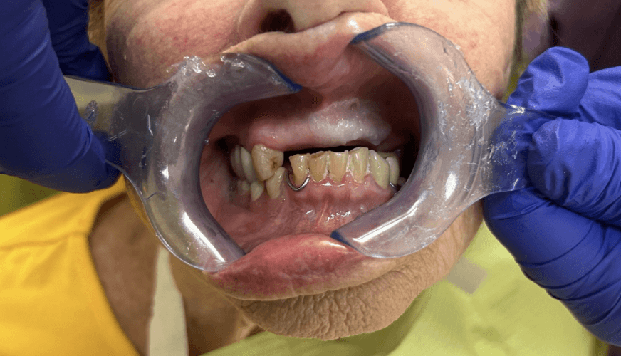
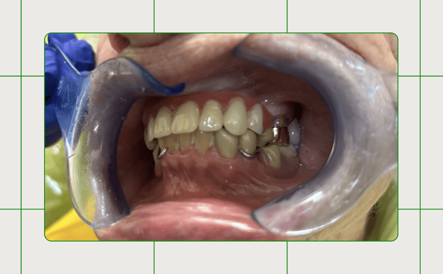
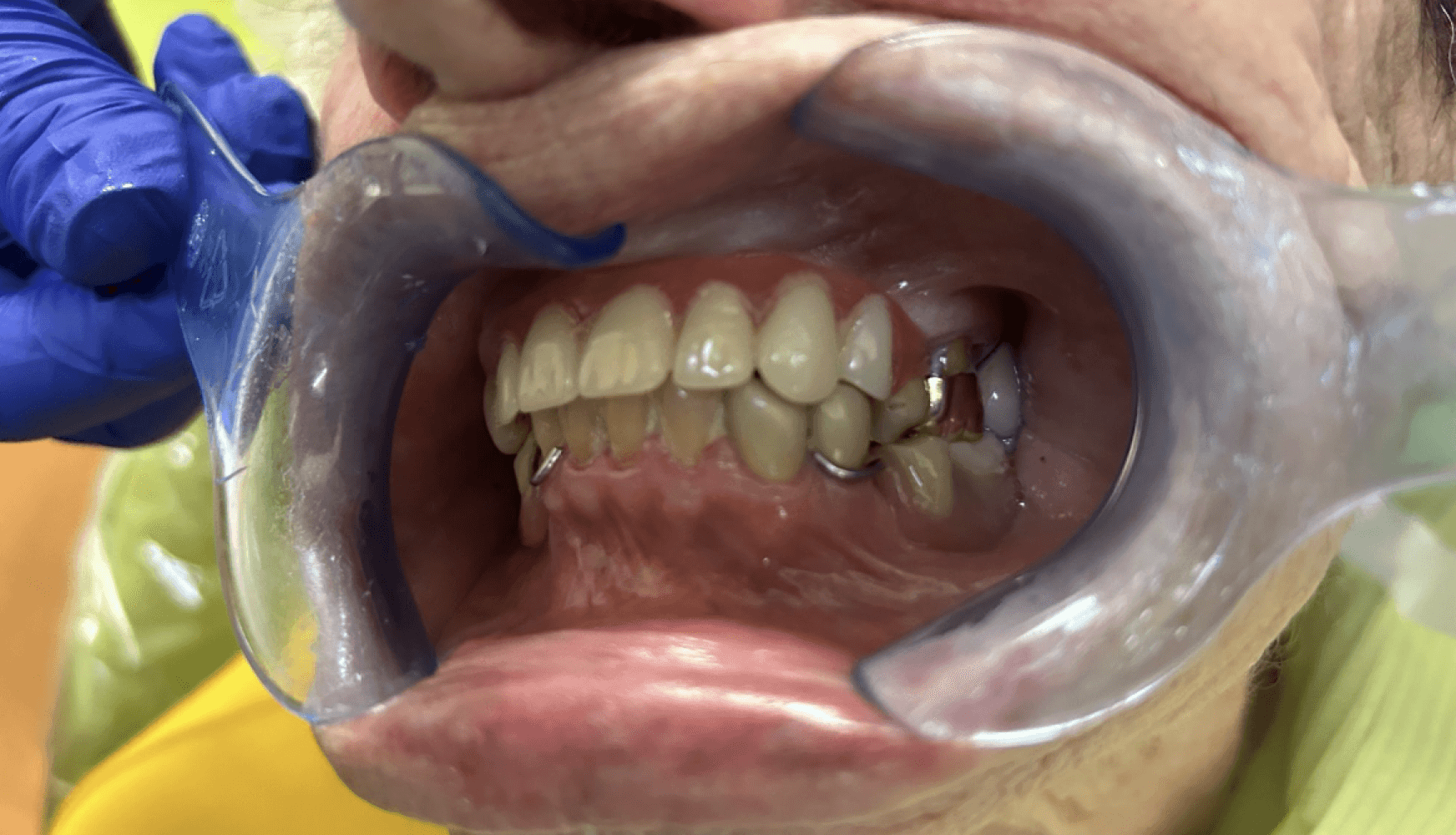
Figure 1:
Figure 1: The patient’s existing dentition prior to replacing her existing maxillary partial.
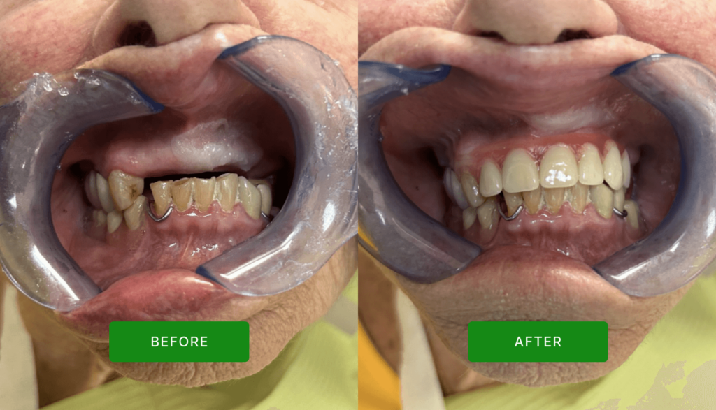
In the case described here, a 90-year-old female-identifying patient presented to the practice stating: “I think I need a new denture, but not one with metal” while pointing to her upper partial. The partial that she requested be remade was an appliance I had delivered in 2021, after extracting teeth #7, 9, and 11. Inspecting the current state of this partial, I could see it was starting to discolor and the teeth were showing signs of wear. The original abutment teeth were still viable to continue to be used as abutments in the remade partial.
Having seen the state of the patient’s three-year-old upper partial and existing abutment teeth, we decided to remake it with a Duraflex partial to restore sites #7-12 accommodating the patient’s requests for palatal coverage and non-metal material.
Case description
Appointment #1:
The patient presented for a prophy and exam, and during this appointment, complained about her current partial. The partial showed signs of discoloration and wear. Given the fact that this partial was replacing anterior teeth, the patient was certain about replacing it. The patient was scheduled for a follow-up appointment to capture all required scans for a new partial a few days later given the time constraints of this initial appointment.
Appointment #2:
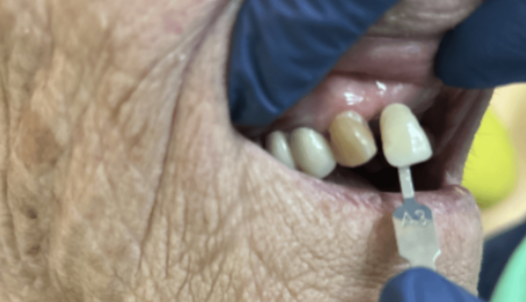
The patient returned for her scanning appointment, and scans (Fig. 2a, 2b) were captured of the upper and lower arches as well as the patient’s bite. The shade of teeth of the future partial was determined to be A3 (Fig. 3) with the Vita Classical shade guide with a TTP (tissue tone pink / light pink) tissue shade base. I opted to order a “try-in with teeth setup” given the aesthetic nature of the partial. I included design notes about incorporating palatal coverage and using non-metal materials, given the patient’s preferences.
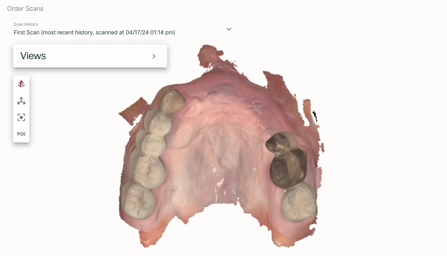
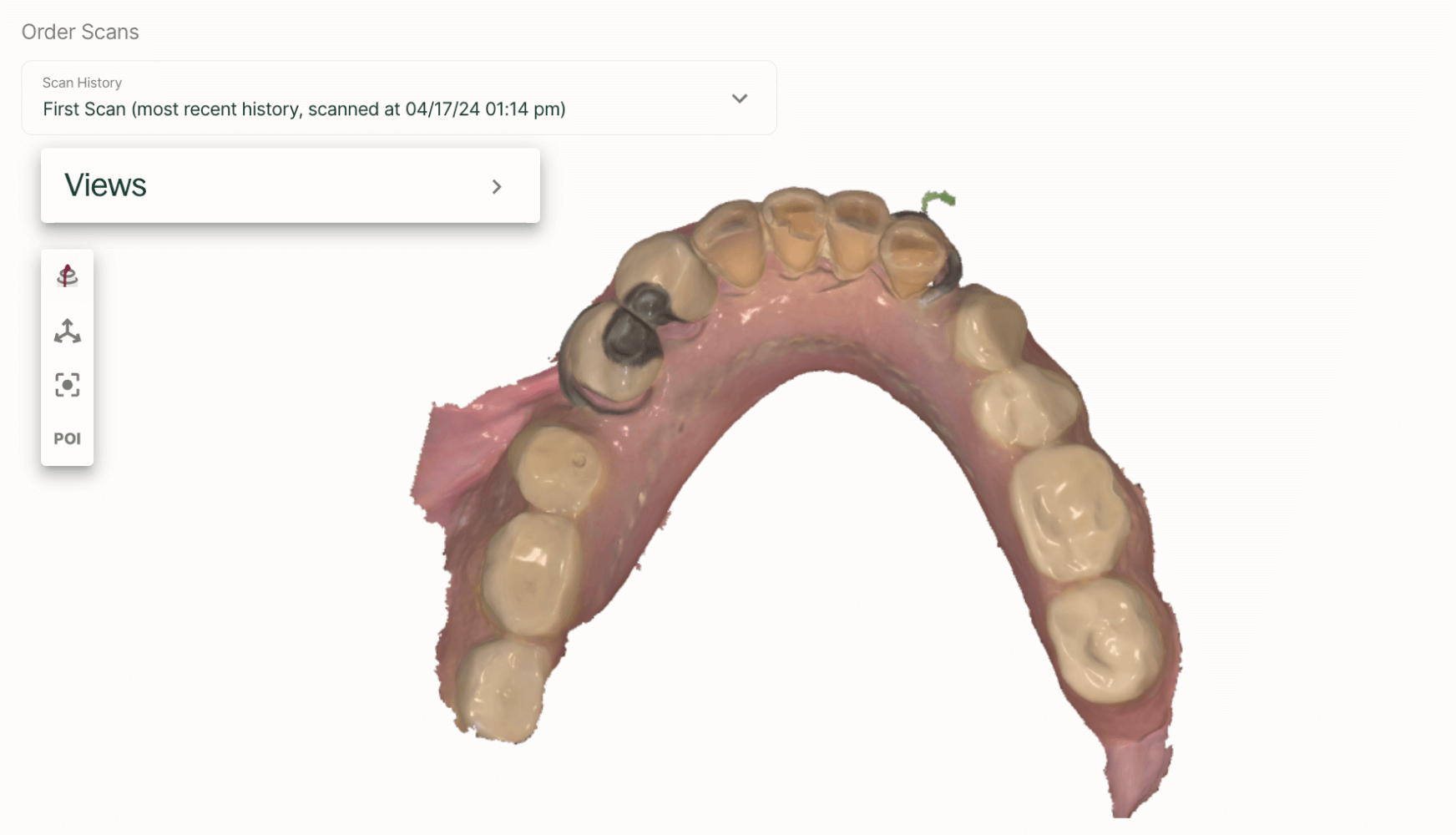
Figures 2a, 2b: Larger pictures of the scans can be found here and here
Figure 2a, 2b: The scans captured during the patient’s scanning appointment. Note the amount of soft tissue included in the scan of the maxillary arch.
Figure 3: A3 was determined to be the agreed-upon shade.
Appointment #3:
During the try-in appointment, the patient’s medical history was reviewed and an extraoral and intraoral exam were performed. The fit of the try-in, its clasps, and its bilateral palatal design were evaluated, and the patient was satisfied with proceeding as is. Aesthetically, the partial base tissue shade, tooth shape, and tooth shade were evaluated with the patient, and the patient was pleased with how the try-in looked. An order was placed for the final partial without any changes to the original design notes and preferences.
Appointment #4:
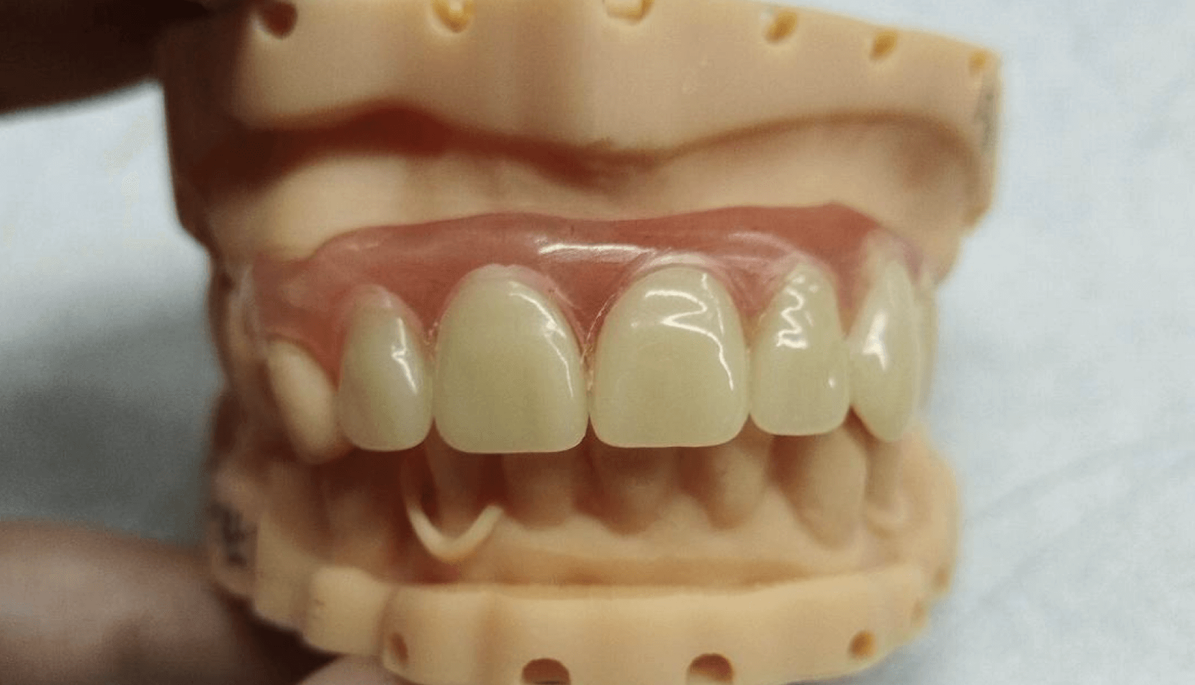
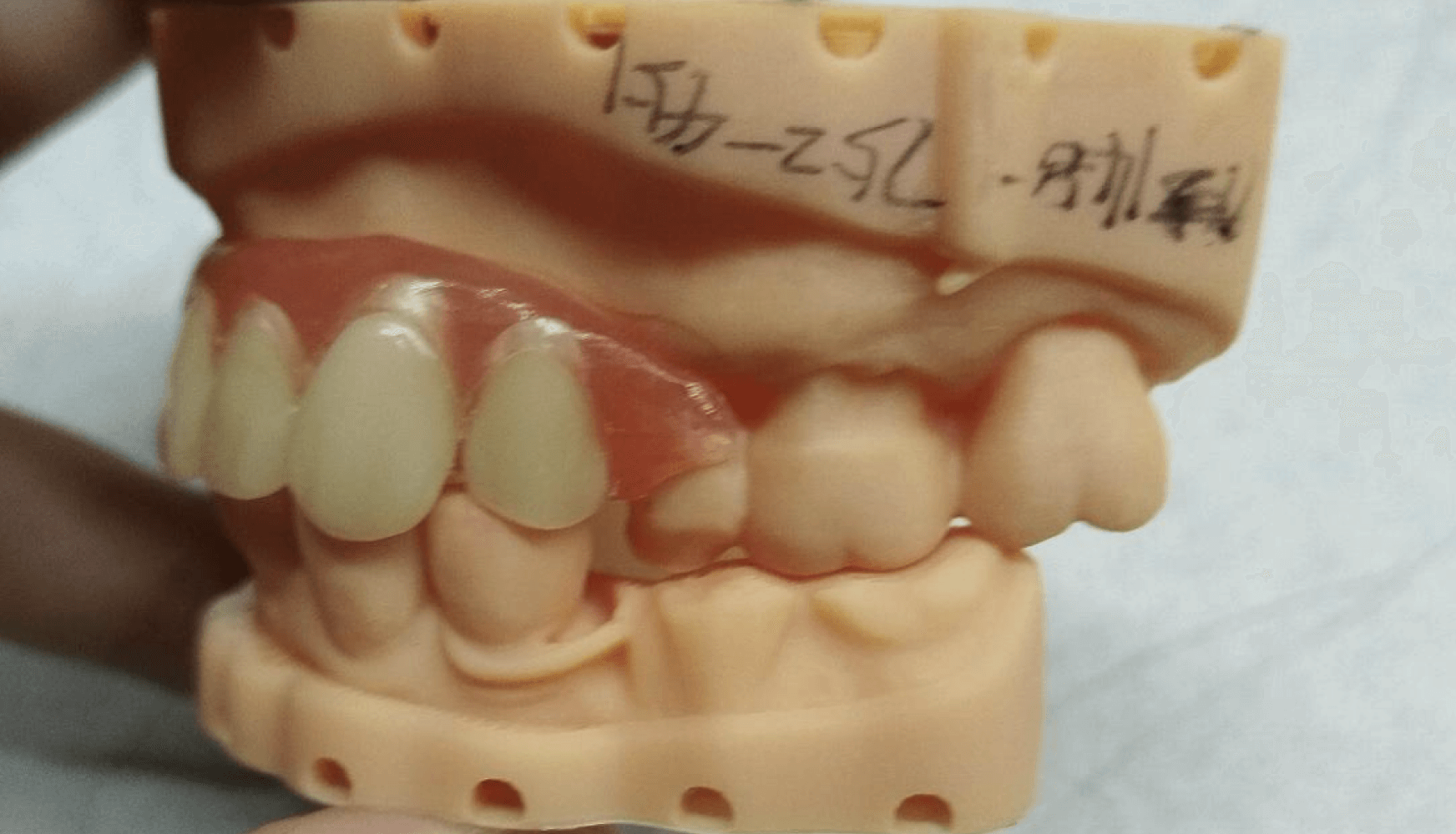
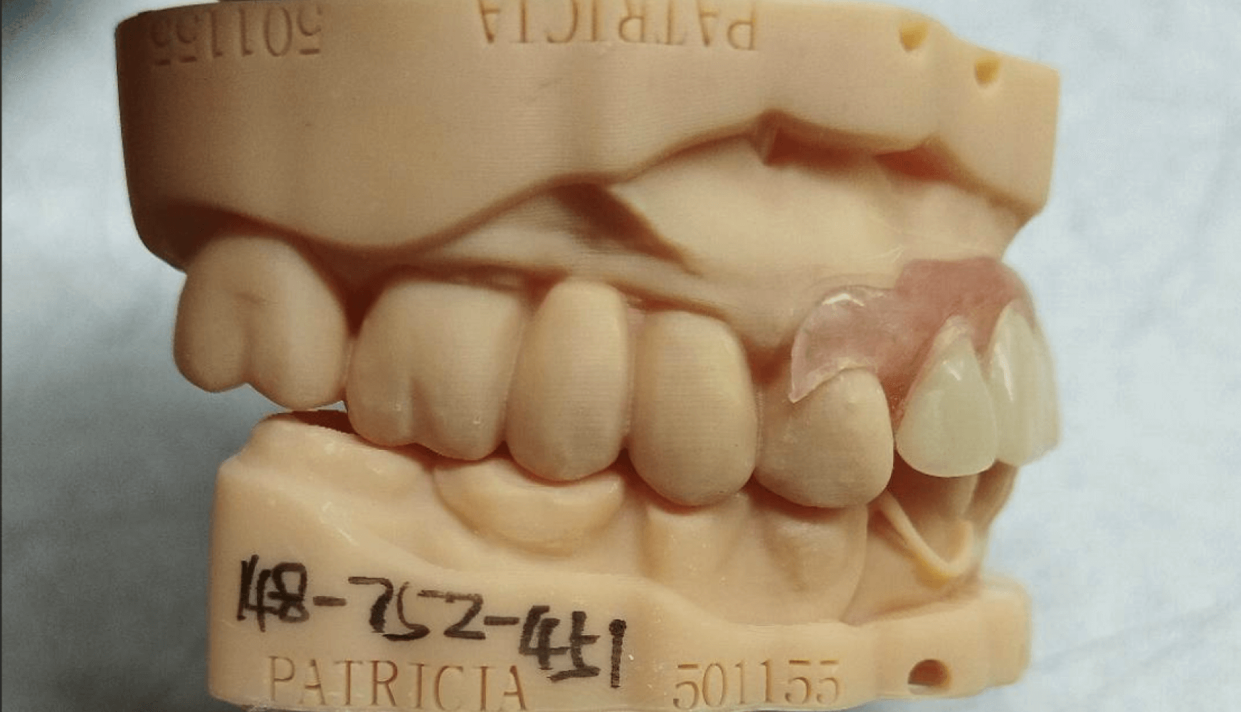
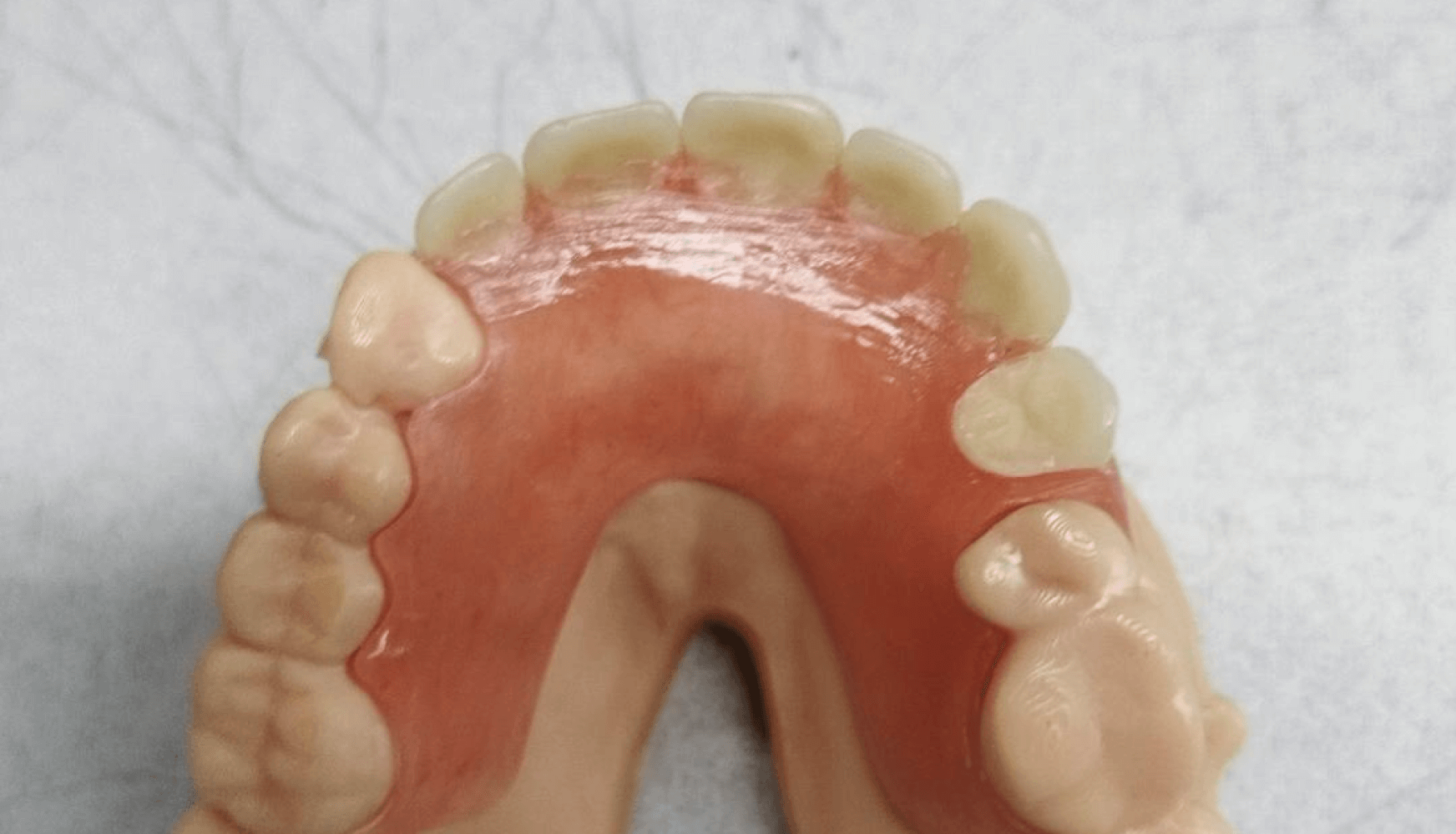
The final partial denture was examined extraorally, and all design specifications were followed (Fig. 4a, 4b, 4c, 4d). The partial was inserted into the patient’s mouth for evaluation, and the patient did not express any pain or general complaints about the feel of the appliance. The patient was instructed to bite, and felt that the bite was slightly incorrect. Minor occlusal adjustment was performed. The partial was reinstated into the patient’s mouth, and the patient expressed that the bite felt correct. The patient was then educated on how to insert and remove the partial. Before dismissal, the patient was reminded that the partial could be adjusted in the future if anything felt uncomfortable or painful in the future.
Figures 4a, 4b, 4c, 4d:
Figure 4a, 4b, 4c, 4d: The fabricated partial denture with its corresponding model. Both tissue shade and tooth shade met the patient’s expectations aesthetically and the overall clasping and fit did so as well.
Appointment #5:
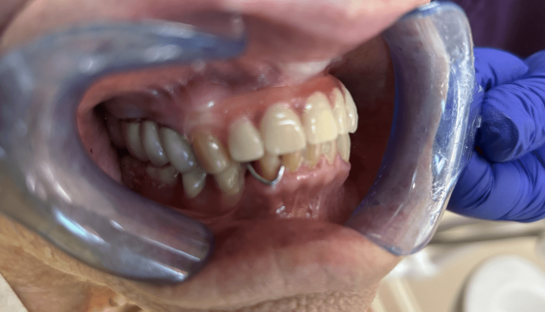
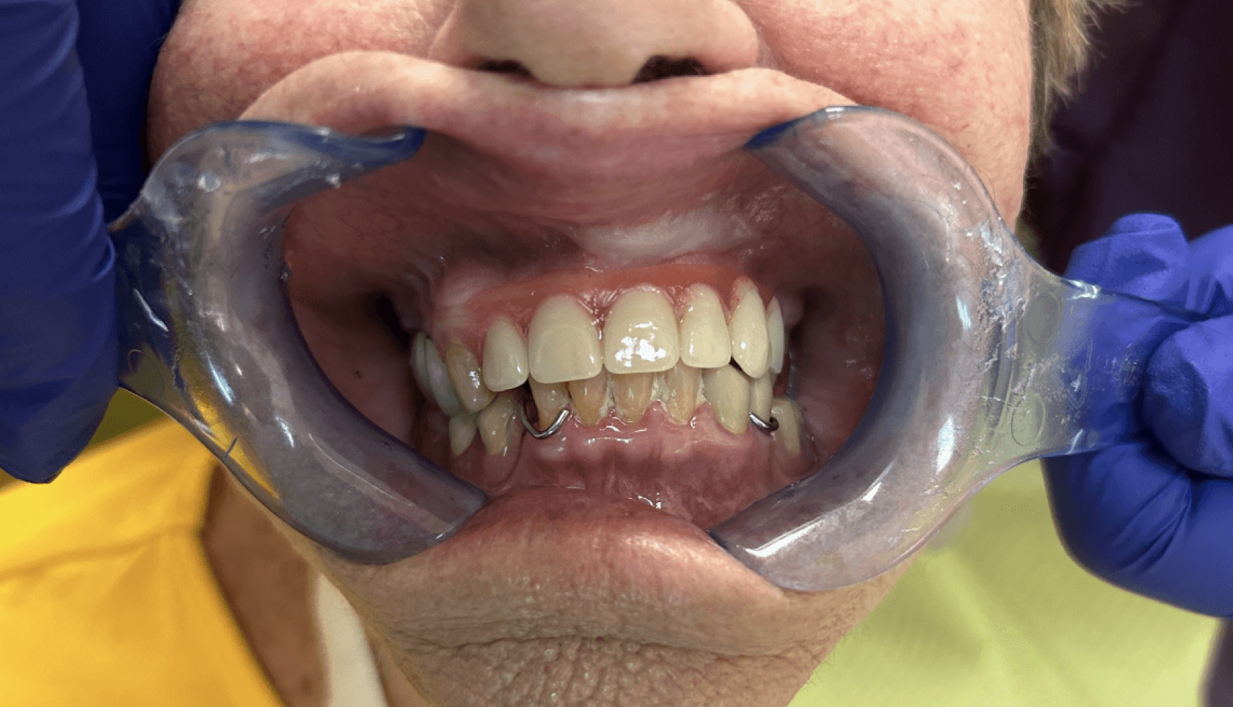
Approximately one week after the delivery of the partial, the patient returned for adjustments, complaining of discomfort, pointing to the vestibule region. The patient also noted that incisors on the partial kept “getting in the way” when chewing and speaking. Upon examination, the labial flange of the partial was causing minor irritation, and the incisors may have been slightly too long. Both issues were easily resolved: the labial flange of the partial was polished and the upper incisors were carefully shortened. The patient left feeling better about the adjustments that were made (Fig 5a, 5b, 5c).
Figures 5a, 5b, 5c:
Figure 5a, 5b, 5c: The maxillary partial denture seated in the patient’s mouth.
Patient before and after
Dandy’s technology
As digital dentistry continues to expand, I always emphasize that technology saves time, and the technology at Dandy does just that. As I was learning to scan for partials and dentures for the first few times, I accessed Dandy’s Help Center to review the overall workflow and read about recommended tips and tricks for edentulous scans. The Help Center gave me video overviews and instructions to get me ready for my very first partial case. It also gave me an easily accessible place to reference this content whenever needed as I was figuring out the techniques that worked for me to capture the scans at the quality that I wanted.
Conclusion & best practices
Scanning soft tissue and edentulous areas requires a different type of touch and finesse compared to scanning teeth. They can be especially challenging for clinicians who are new to digital dentures, but shouldn’t be a deterrent—soft tissue and edentulous scans come with their own learning curve. When performing this type of scan, I always focus on saliva control and keeping my scanner as close to the gingiva as I can. It is critical to not rush when scanning; I’ve found that scanning too quickly actually works against me and prolongs the scanning process. If possible, I start my scans by beginning over the occlusal surface of a tooth if available. Then, I slowly slide the scanner towards the soft tissue, lowering the scanner closer to the edentulous site as I move from tooth-to-tissue. As I scan, I will sometimes perform a slow, gentle rock by rotating my wrist, helping the scanner find more reference points along the soft tissue. Ultimately, a slower pace, saliva control, and a little bit of patience make these scans achievable and give Dandy the imaging they need to design partials.
About the author
Dr. Natalia Elson is a Clinical Assistant Professor at NYU Dentistry and Clinical Assistant Professor at Stony Brook School of Dental Medicine. She graduated from Dnepropetrovsk Medical Institute, School of Dental Medicine in 1977 and from NYU College of Dentistry in 2010.
She also finished General Practice Residency in Ukraine in 1978, Residency in Oral Surgery in Georgia in 1986 and General Practice Residency in Stony Brook University in 2011. Her family moved to the US from Estonia in 2003 where she was running private practice and lecturing at Tartu University. Dr. Elson opened her private practice in Islip , NY in 2012. Dr. Elson has publications in peer-reviewed journals in Estonia and the United States, lecturing in different dental events and to students at NYU Dentistry.

