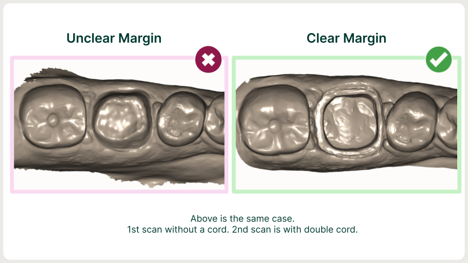
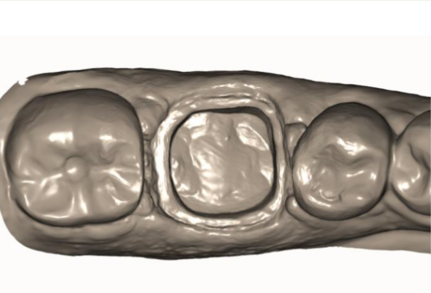
We love the double cord retraction technique in dentistry for a number of reasons. Simply put, it’s better than dentists using a single packing cord dental technique when they’re doing an impression-taking technique for replacement and restoration of teeth with artificial substitutes. The double cord method is particularly helpful for crown and bridge restorations.
You might need a refresher, ‘what is the double cord technique?’
The double cord technique is a dental procedure that involves placing packing cord between the tissue and the tooth when taking prosthetic impressions. One cord goes deep and one is shallow.
The dentist needs to displace the gingiva in order to obtain an accurate impression for a fixed prosthesis. Accuracy of gingival displacement using the retraction cord dental is particularly important when the finish line is at or within the gingival sulcus.
This gingival cord retraction technique allows for more significant retraction of the gums.
Often, a dentist can pull out the second cord (which would be closer to the margin and shallower) and keep the first cord (which would be deeper) in place if it sits below the margin for a final impression scan.
By using double cord as part of the dental impression technique, your margins are better when it comes to getting an accurate impression. Packing cord dental practices with two cords can help prevent tissue collapse when getting the impression so you’re able to do the best prosthetic impression possible without worrying about a single dental cord tearing.
Here, learn more about how the double cord retraction technique is best suited for margins when taking digital impressions and how an intraoral scanner can help.
Dandy offers dental practices a free intraoral scanner.
The difference between single cord and double cord retraction techniques
Retraction cord can be thought of as a piece of thin string that gets placed in between the crown-prepped tooth and the surrounding gums. Retraction cord sometimes can be packed beneath the margin of the prepped tooth, but sometimes, it is packed at nearly the same level as the margin. The depth usually depends on the patient’s gums on a case-by-case basis.
If the dentist “pulls the cord,” the gums do not suddenly snap back into place and cover the margin. Instead, the gums very slowly and gradually return to their original position. When a dentist pulls the cord, he/she must move quickly to scan their final impression before the gums cover the margin of the tooth again.
Still, polyvinyl siloxane (PVS) impressions result in a high-likelihood of pulls/distortions and a high refabrication rate. That’s not an ideal situation for you or your patient.
A single cord is an adequate option of tissue retraction as long as the margin is visible and the tissue has not collapsed prior to scanning.
If you’re following the single cord retraction technique, follow these steps:
- Ensure the retraction cord dental is soaked in hemostatic material.
- Leave it in place for 5 minutes.
- Then pull the cord and scan for optimal results.
If you’re using the double cord technique—our preferred one!—follow these steps:
- Pack two knitted cords—soaked in hempstatic material and at different diameters—into the sulcus.
- Leave the larger cord in place for at least five minutes before pulling.
- Often, a dentist can pull out the second cord (which would be closer to the margin and shallower) and keep the first cord (which would be deeper) in place if it sits below the margin for a final impression scan.
- Blast with air to ensure prep and tissue surrounding prep is dry.
- Take the impression using an intraoral scanner.
- The dental cord can stay in during the final impression scan if the cord is packed below the margin, and is packed deep enough so that none of the cord is covering the margin.

How digital dentistry changed the technique for packing dental cords
Taking a PVS impression before the age of digital scanners provided physical retraction on the tissue enough to flow in between the margin and the free gingiva. This allowed for all prep styles to be implemented by dentists. Despite this benefit, PVS impressions result in a high-likelihood of pulls/distortions and a high refabrication rate.
Use an intraoral scanner to minimize the chances of a refabrication rate during a cord retraction technique.
- Digital scans benefit most from shoulder or chamfer prep styles—overall we prefer shoulder style margin prep for its visibility.
- When prepping with a shoulder, the margin is clearly visible on the scan—ensuring the restoration matches perfectly to the finish-line. In clinical situations where this is not possible, a medium to heavy chamfer style of margin will work.
- Keep in mind, the ideal occlusal space should be at least 1-1.5mm
- For optimal scanning results apply the Double Cord Technique (available in sizes 000, 00, 0, 1, and 2) due to the increased accessibility and improved displacement of tissue.
Another benefit to using an intraoral scanner for a digital impression? If you see that the scan isn’t ideal, minimal effort is required to re-scan for optimal results.
Producing better impressions with double cord retraction
As your digital dental partner, we want to produce the highest quality prosthetics and restorations for our doctors. The first step to a successful restoration is an accurate digital impression and clear margins. Whether you’re prepping a zirconia crown, prepping for veneers, or prepping for another crown or bridge restoration, gingival cord retraction using the double cord technique is what we recommend to get consistent results, every time.


